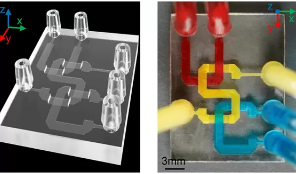
Engineers fabricate microfluidic chips for biomedical applications
Microfluidic devices have important applications in medical research, drug development and diagnostic tools. The compact devices have channels carved into a resin chip that enable testing on a microscale and are labor-intensive to produce.
A team of engineers based at the University of Southern California, pursuing U.S. National Science Foundation-supported research, developed a specialized 3D-printing technique that allows microfluidic channels to be fabricated on chips at a precise microscale not previously achieved.
The team used vat photopolymerization, a type of 3D-printing technology that harnesses light to manage the conversion of liquid resin material into a solid form.
"This is the first time we've been able to print something where the channel height is at the 10-micron level and we can control it accurately, to an error of plus or minus one micron," said Yong Chen, one of the authors of the study.
The process involves a using liquid photopolymer resin to construct the object layer by layer and curing each layer with ultraviolet light. During fabrication, the printed object is moved to build additional layers.
Chen said, "When you project the light, ideally, you only want to cure one layer of the channel wall and leave the liquid resin inside the channel untouched, but it's hard to control the curing depth, as we are trying to target something that is only a 10-micron gap."
The team created channels by using a platform that moves between the light source and the printed object to block the light from curing the liquid resin within the channel. After the curing process the residual liquid resin is cleared from the channel.
"There are so many applications for microfluidic channels,” Chen said. “You can flow a blood sample through the channel, mixing it with other chemicals, so you can, for example, detect whether you have COVID or high blood sugar levels.”
Right now, said Chen, “we use biopsies to check for cancer cells, cutting organ or tissue from a patient to reveal a mix of healthy cells and tumor cells. Instead, we could use simple microfluidic devices to flow the sample through channels with accurately printed heights to separate cells into different sizes, so we don't allow those healthy cells to interfere with our detection."
